

EExploring Phagocytosis and Macropinocytosis

Nobukazu Araki, Professor
Histology and Cell Biology, Kagawa University School of Medicine
Japan
Original Movies
* Original Promotion Video by Araki lab *
Digest of live cell imaging project exploring phagocytosis and macropinocytosis
Movie1. Phagocytosis of IgG-erythrocytes
Phagocytosis of IgG-opsonized sheep erythrocytes by bonemarrow-derived
macrophages.
Time-lapse images of phase-contrast microscopy were taken at 10-second
intervals.
Movie speed x60 real time.
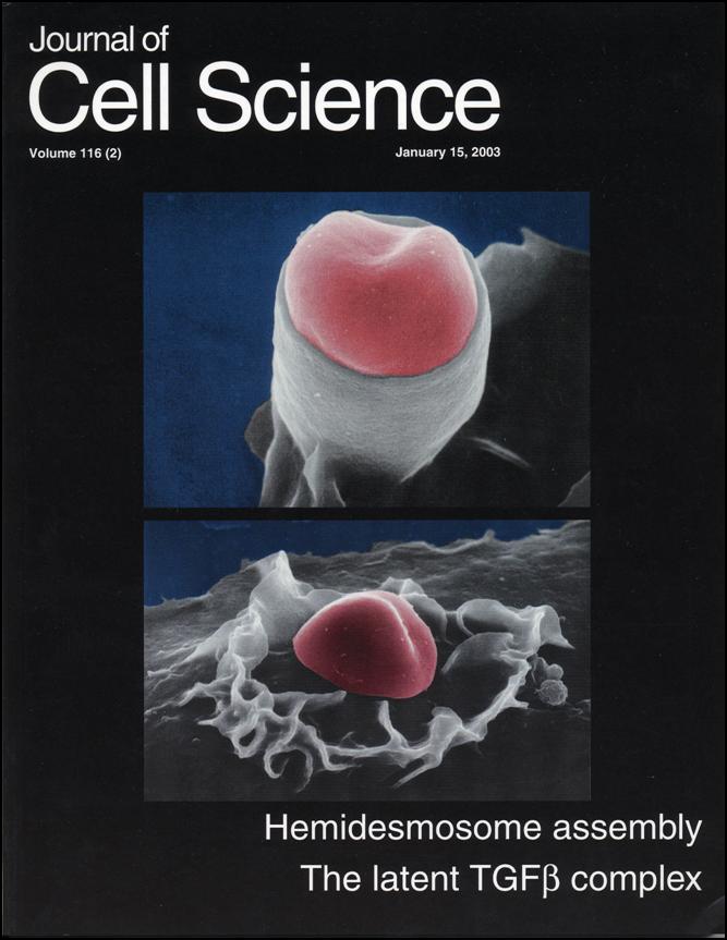
Movie 2. Actin Dynamics during FcR-mediated Phagocytosis
Fc receptor-mediated phagocytosis of IgG-erythrocytes by RAW264 macrophages
expressing GFP-actin.
Pseudopod extension around IgG-erythrocytes is driven by actin-polymerization.
Note the phagocytic cup squeezing IgG-erythrocytes.
Actin filaments are dissassembled from the membrane of basal portion of
phagocytic cups.
After closure of phagocytic cups, few actin filaments are associated with
intracellular phagosomes.
Ref J. Cell Science 2003 Araki et al.
Movie 3. Movement of peroxisomes during phagocytosis
Time-lapse movie of RAW264 macrophages expressing GFP-fused peroxisomal proteins.
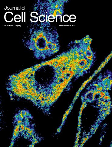
Movie 4. Changes in PI(4,5)P2 and PI(3,4,5)P3 levels during macropinocytosis
in EGF-stimulated A431 cells
A431 cells co-expressing EGFYFP-Akt-PH domain (red:PI(3,4,5)P2) and CFP-PLC PH domain (green: PI(4,5)P2)
were stimulated with EGF to induce ruffling and macropinocytosis.
Ref Exp.@Cell Res. 2008 Araki et al.
Movie 5. Changes in PI(3,4,5)P3 and actin during macropinocytosis in EGF-stimulated A431 cells
A431 cells co-expressing CFP-Akt-PH domain (green) ÆGFP-actin (red) were
stimulated with EGF.
F-actin was dissassembled from the macropinosome membrane when PI(3,4,5)P3
levels was becoming high.
Exp.@Cell Res. 2008 Araki et al.
Link to published movies
Movie 6. Live cell imaging of PtdIns(3)P by expressing YFP-2~FYVE in control A431 cells.
Time-lapse images of A431 cells expressing YFP-2~FYVE were acquired at 10 second intervals by digital fluorescence microscopy using MetaMorph imaging system and assembled into a QuickTime movie at a frame rate of 5 fps. Cell structure and Finction 2007 Araki et al
Movie 7. Enlarged time-lapse movie of the selected area from Movie 6 showing
the dynamics of YFP-2~FYVE-positive macropinoses
Time-lapse images of A431 cells expressing YFP-2~FYVE were acquired at 10 second intervals by digital fluorescence microscopy using MetaMorph imaging system and assembled into a QuickTime movie at a frame rate of 5 fps. Cell structure and Finction 2007 Araki et al
.
Movie 8. Time-lapse movie showing the inhibition of PtdIns(3)P production
by 3-MA treatment.
Cells expressing YFP-2~FYVE were treated with 3-MA for 15 min and stimulated with EGF. Macropinosome formation was apparent, but the recruitment of YFP-2~FYVE to the macropinosomes was abolished by the 3-MA treatment.
Movie 9. Contractile activity of phagicytic cups published in Jounal of
Cell Science
QuickTime Video
Movie 10. Dynamics of Rab21 in macrophages during macropinocytosis
Time-lapse movie showing the localization of GFP-Rab21 in RAW264 cells. Note the association of Rab21 with macropinosomes in the M-CSF-stimulated RAW264 cell. The images were collected at 10-s intervals. This is a representative of five cells, and similar results were obtained from three-independent experiments. (2.55 MB MOV) Other moives are available in Open Acess Journal PLoS ONE 2009 Egami and Araki